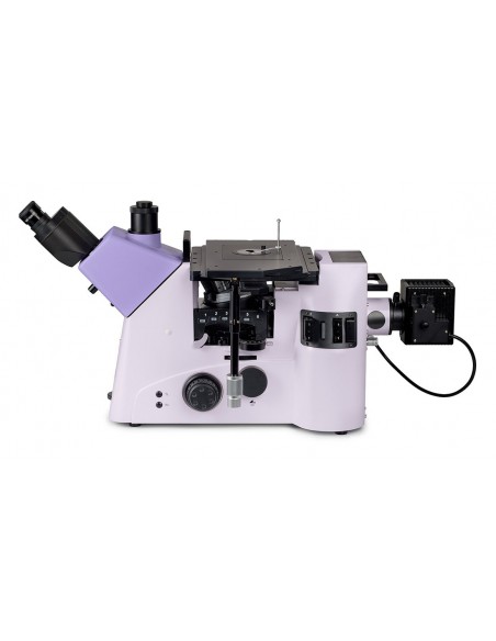
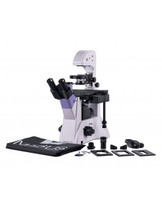
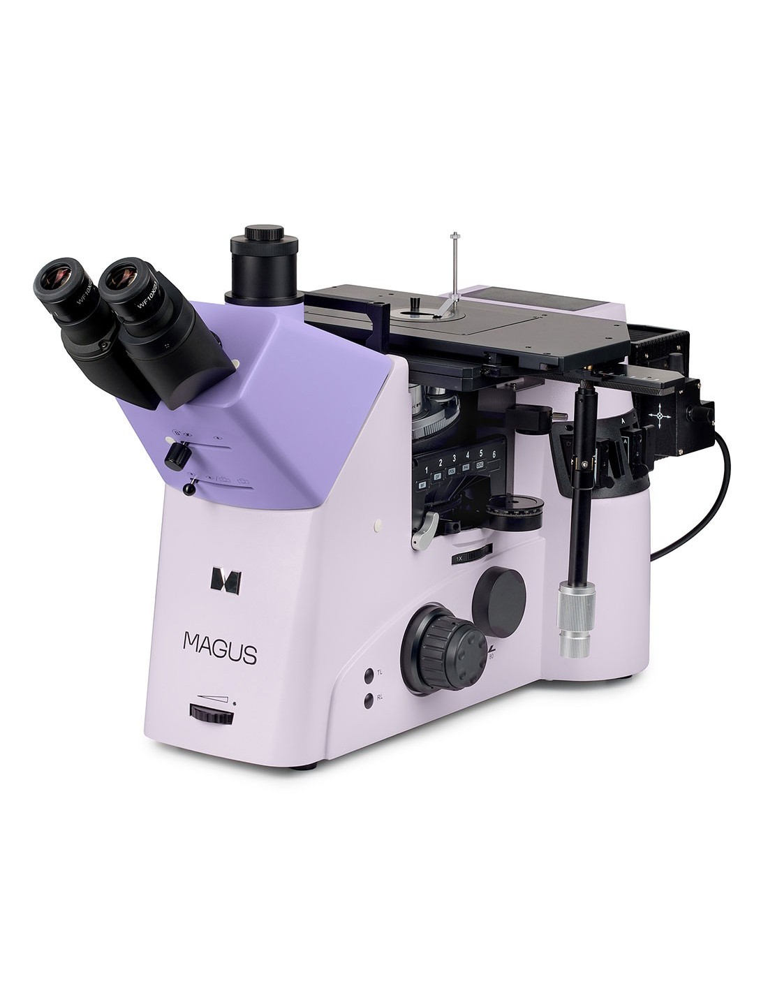
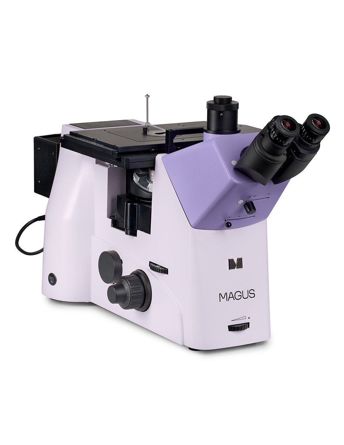
This microscope is designed for studying the microstructures of metals, alloys, semiconductor materials and other opaque samples.
The inverted microscope design does not limit the size of the sample examined; only its weight is limited up to 30 kg. The sample to be analyzed must first be appropriately processed, then positioned on the stage with the surface of interest facing downwards.
This microscope can study opaque objects in reflected light using bright field, dark field and polarization microscopy techniques and differential interference contrast.
DIC is an advanced polarization contrast technique. The method increases the depth of field and clarity of the image, provides additional information about the structure of the studied object and displays irregularities on the surfaces studied with color. Flat surfaces are colored the same way, but a surface with even minimal differences in height will have a non-uniform color. The object of study will appear in relief.
The microscope can be used in various fields (metallurgy, engineering, aerospace, nuclear and energy sectors) as well as in research laboratories and technical universities.
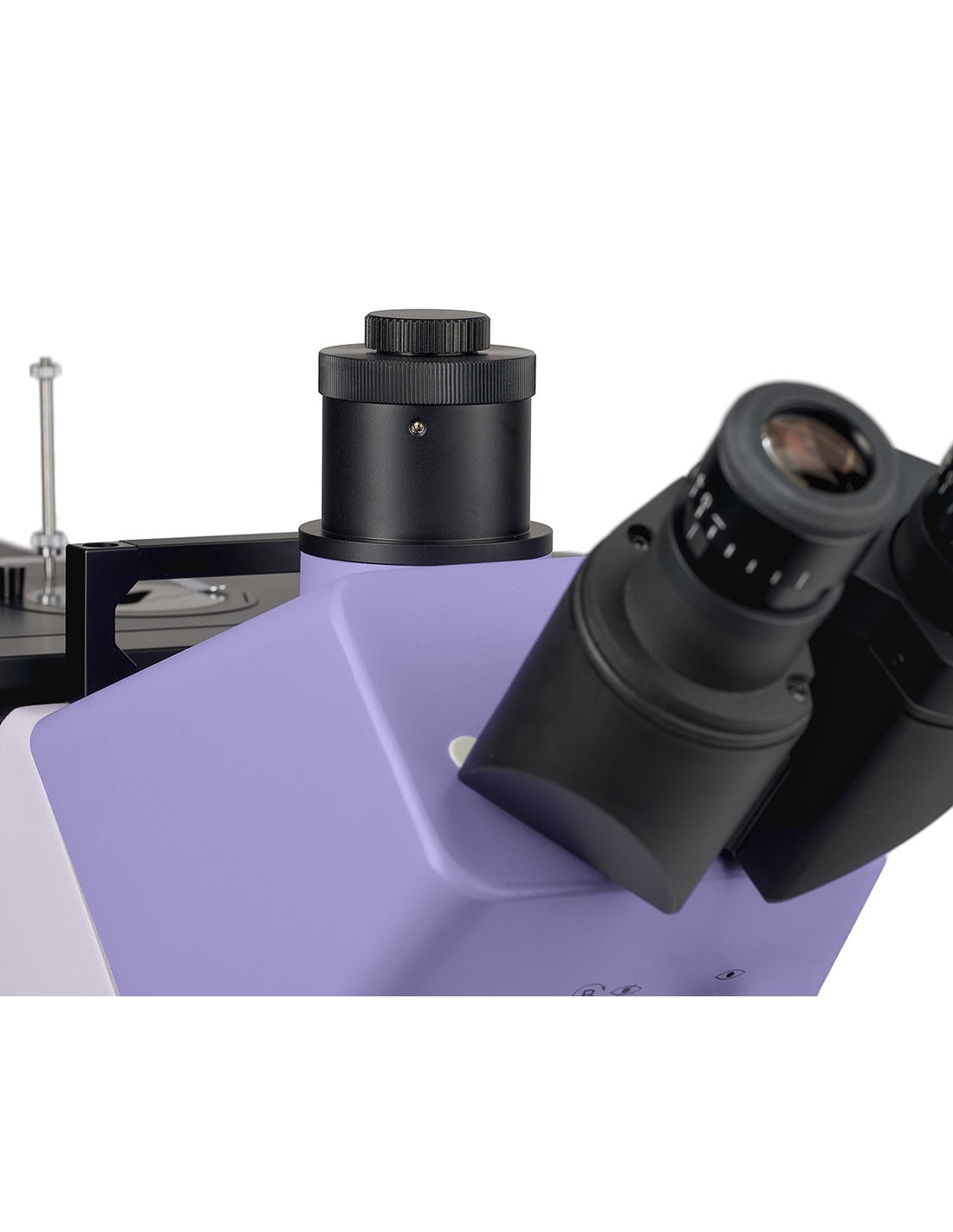
Microscope head
Trinocular head with infinity corrected optics. The 45° tilt angle offers comfortable conditions for the researcher to work while sitting or standing. The user can assume a natural pose, therefore, fatigue does not accumulate during the working day.
A Bertrand lens is incorporated into the microscope head; a regulator on the front of the muzzle is used for insertion into the optical system. Bertrand lenses are used for cognitivist studies.
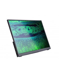
The digital camera is installed in the trinocular tube or in one of the side imaging outlets on the body of the microscope. The beam on the trinocular head can be split 100/0, 50/50 or 0/100. The beam split on the right photo output is 100/0 and 20/80; on the left is 100/0 and 0/100.
The basic package includes 10x/23 mm eyepieces with diopter adjustment and long eye relief for working with glasses.
Nosepiece revolver
strong>
The nosepiece nosepiece with 5 objectives is installed on the stand, below the stage.
The differential interference contrast method is implemented using the DIC sliders: one is used with low magnification and the other with medium and high magnification objectives. The DIC slider is installed in a special housing of the objective nosepiece.
Turret for installing contrast modules
Under the objective nosepiece on the microscope stand , there is a turret to install contrast modules. Six modules can be installed. Rotating the turret changes the observation mode and switching from one contrast method to another occurs quickly and without complex settings.
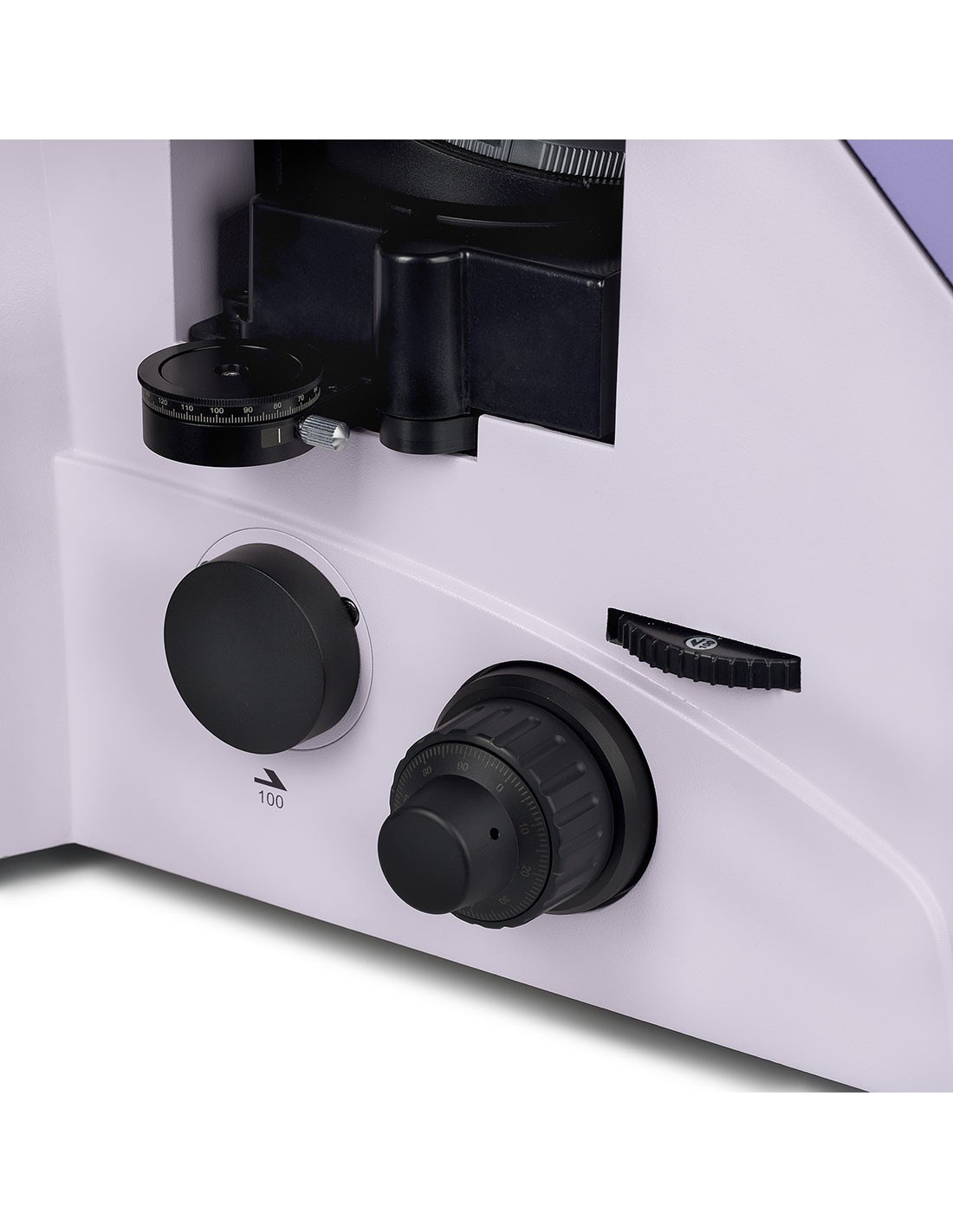
Switch with additional magnifying glass
A rotary switch is used to insert the 1x main lens or an additional 1.5x magnifying glass into the optical path. A magnifier increases the magnification of a microscope without purchasing or installing additional accessories. The switch is located on the right side of the microscope body, under the nosepiece.
Objectives
Plan semi-apochromatic and plan apochromatic objectives with a long working distance that are designed for brightfield and darkfield microscopy techniques. They provide better correction of chromatic and spherical aberration than planachromatic lenses.
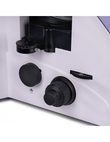
Focusing mechanism
The coaxial fine focus knobs ( micrometric screw) and coarse screw (macrometric screw) are positioned on both sides at the base of the microscope. The fine focus handle on the right has special finger grooves, and the left one has a scale.
The coarse focus lock knob helps to quickly adjust the microscope after changing the study object. The knob is located on the left side of the microscope on the same axis as the focusing mechanism.
The ring on the right side adjusts the tension of the coarse focus screw. The user adjusts the right tension for the observations.
Stage
The object is moved by moving the stage along two axes. An insert with a hole of a suitable diameter is installed in the center of the table. A sample holder/slide holder system is used to hold the observed object in place.
The three-layer coating protects the stage from scratches and other surface damage when working with large, heavy specimens.
The long control handle of the tilting table ensures user comfort during work: the hand rests on the table effortlessly. The handle can be installed on the right or left side of the coffee table.
Light source
The lamp holder has a 100W halogen bulb. It is bright enough for work on objectives with magnifications from 4x to 100x, not only in bright field and polarized light, but also in dark field microscopy techniques. The halogen lamp emits light with a color temperature that guarantees greater comfort for the eyes.
Reflected light lighting
The lighting system makes it possible to Köhler's lighting setup. Aperture and field diaphragms are centered by default and do not require further adjustment. If necessary, the diaphragms can be adjusted using the centering screws.
The analyzer and polarizer are for the polarization microscopy technique. The polarizer is fixed and the analyzer rotates 360°.
A series of light filters helps regulate color reproduction.
Ergonomic design
Physical discomfort causes fatigue and reduces productivity. The ergonomic design of the microscope plays an important role in daily scientific research.
MAGUS Metal V790 DIC provides comfort to the user while working.
The microscope is operated with minimal hand movement as the long handle stage control and fine focus knob are located in the same working area.
The focus knobs are located on the bottom of the body. The user does not strain his hands. Thanks to the smooth movement of the mechanism, the user can concentrate on the object effortlessly.
Key features:
- Suitable for studying opaque dimensional objects up to 30 kg
- The Bertrand lens for Conopia and Orthoscopy
- An additional magnifying glass increases the magnification of the microscope by 1.5x; is inserted into the optical system via a switch on the microscope head
- Three independent options for mounting a digital camera and a monitor: a trinocular tube and two side tubes on the body; beam division on the trinocular head: 100/0, 50/50, 0/100, on the body: 100/0 and 20/80 (right photo output), 100/0 and 0/100 (left photo output)
- Plan semi-apochromatic and plan apochromatic objectives with extended working distance are used for work in brightfield and darkfield techniques
- Multifunctional turret with 6 slots for installing modules of different contrast methods
- Reflected light halogen illuminator with increased power of 100 W, specific for the dark field method
- Köhler illumination, centered elements of the illumination system, dark field techniques , bright field, polarization and DIC
- Color filters reduce the intensity of certain wavelengths and adjust the color reproduction in microscope photography
- Large stage with inserts and long control handle for comfortable operation
The package contains:
- Stand with integrated power supply, setting mechanism focus, stage, nosepiece revolver and turret to install contrast modules
- Lamp housing
- Trinocular head with Bertrand lens
- Semi-apochromatic plan objective at infinity : Plan S-Apo BD 5x/0.15 ∞/- WD 20 mm
- Plan semi-apochromatic infinity objective: Plan S-Apo BD 10x/0.30 ∞/- WD 11 mm
- Plan semi-apochromatic infinity objective: Plan S-Apo BD 20x/0.45 ∞/0 WD 3.1 mm
- Plan apochromatic infinity objective: Plan Apo BD 50x/0.80 ∞/0 WD 1 mm
- Infinity plan apochromatic objective: Plan Apo BD 100x/0.90 ∞/0 WD 1 mm
- 10x/23 eyepiece mm with long eye relief and diopter adjustment (2 pcs.)
- Eyecup (2 pcs.)
- DIC slider for lenses (2 pcs.)
- Polarizer tool
- Filters set
- C-mount adapter
- AC mains power cord
- Dust cover < li>User manual and warranty certificate
Available on request:
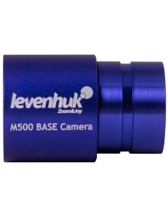
- Digital camera
- Cover glass calibration
| Brand | MAGUS |
| Warranty | 5 лет |
| EAN | 5905555019512 |
| Package size (LxWxH) | 76x61x58 cm |
| Shipping weight | 34.9 kg |
| Type | light/optical, metallographic |
| Head | trinocular |
| Vertical tube | Siedentopf, with Bertrand lens, beam division 100/0, 50/50, 0/100 |
| Head inclination angle | 45° |
| Magnification, x | 50–1000 in basic configuration (50–1500x with additional integrated 1.5x magnifying glass) |
| Magnification range | 800x to 1280x |
| Eyepiece tube diameter, mm | 30 |
| Eyepieces | 10x/23 mm, long eye relief (2 pcs.) |
| Objectives | plan semi-apochromatic and plan apochromatic objectives, at infinity: 5x/0.15; 10x/0.30; 20x/0.45; 50xs/0.80; 100xs/0.90; parfocal height 45 mm |
| Nosepiece revolver | for 6 objectives, with DIC housing |
| Opening distance work, mm | 20 (5x); 11 (10x); 3.1 (20x); 1 (50xs); 1 (100xs) |
| Interpupillary distance, mm | 48 — 75 |
| Stage, mm< /td> | 340x230 |
| Range of movement of the stage, mm | 130/85 |
| two-axis, three-layer mechanics, with coaxial X/Y handle with low position and tilt capacity, maximum load 30 kg, table inserts |
| Eyepiece diopter adjustment, diopters | ±5D on each eyepiece |
| Eyepiece diopter adjustment | yes |
| Diaphragm | adjustable aperture diaphragm, field diaphragm with adjustable iris |
| Focus |
| td> | coaxial, coarse (10 mm, with fixing knob and micrometer screw tension adjustment knob) and fine (0.002 mm micrometer screw, 0.2 mm/rev) |
| Lighting | halogen |
| Brightness adjustment | yes |
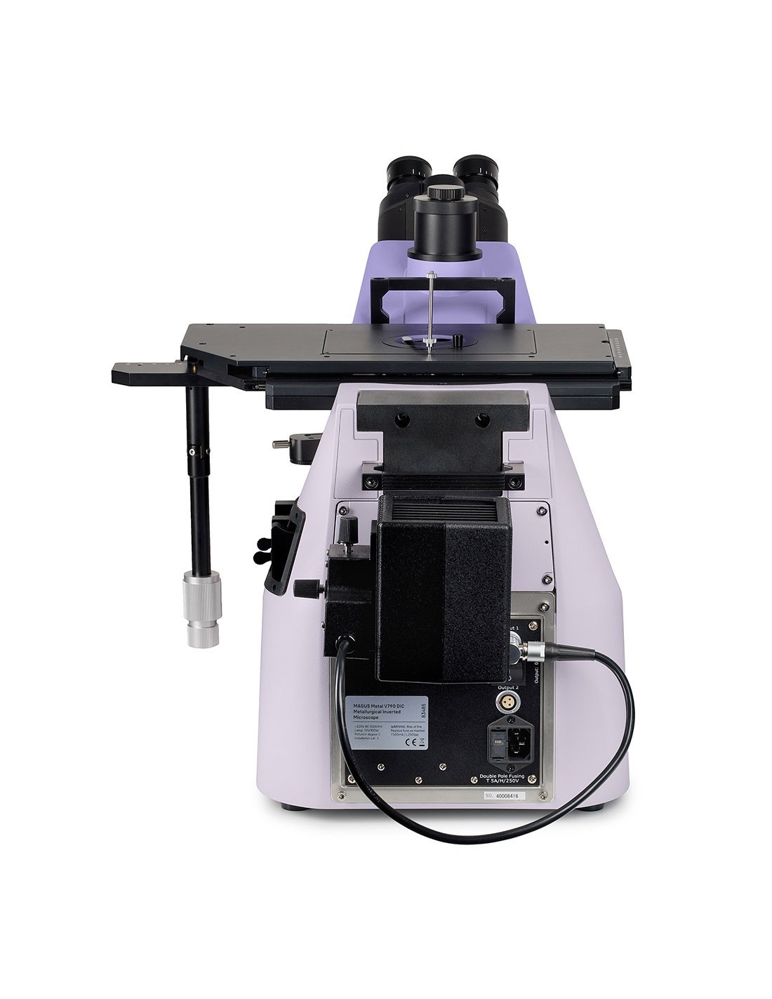
| Power supply | AC mains, 220±22 V, 50 Hz |
| Type of light source | 12 V/ 100 W |
| Optical filters | yes |
| Operating temperature range, °C | 5 — 40 |
| Extra elements | polarizer and analyzer, DIC slider for objectives, turret for installing contrast modules |
| User level | expert users, professionals |
| Assembly and installation difficulty level |
| Application | metallographic |
| Lighting position | upper |
| Research method | bright field, dark field, polarized light, DIC |
| Set with pencil case/case/bag | anti-dust cover |


