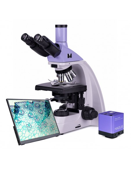
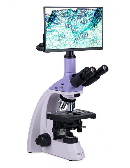
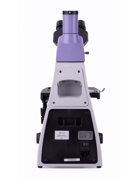
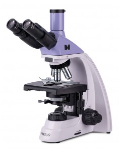
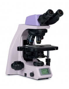
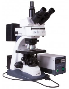
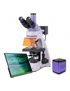
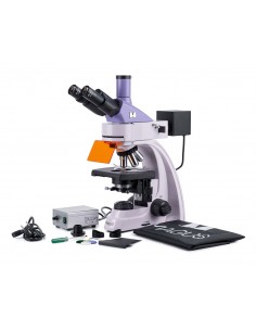
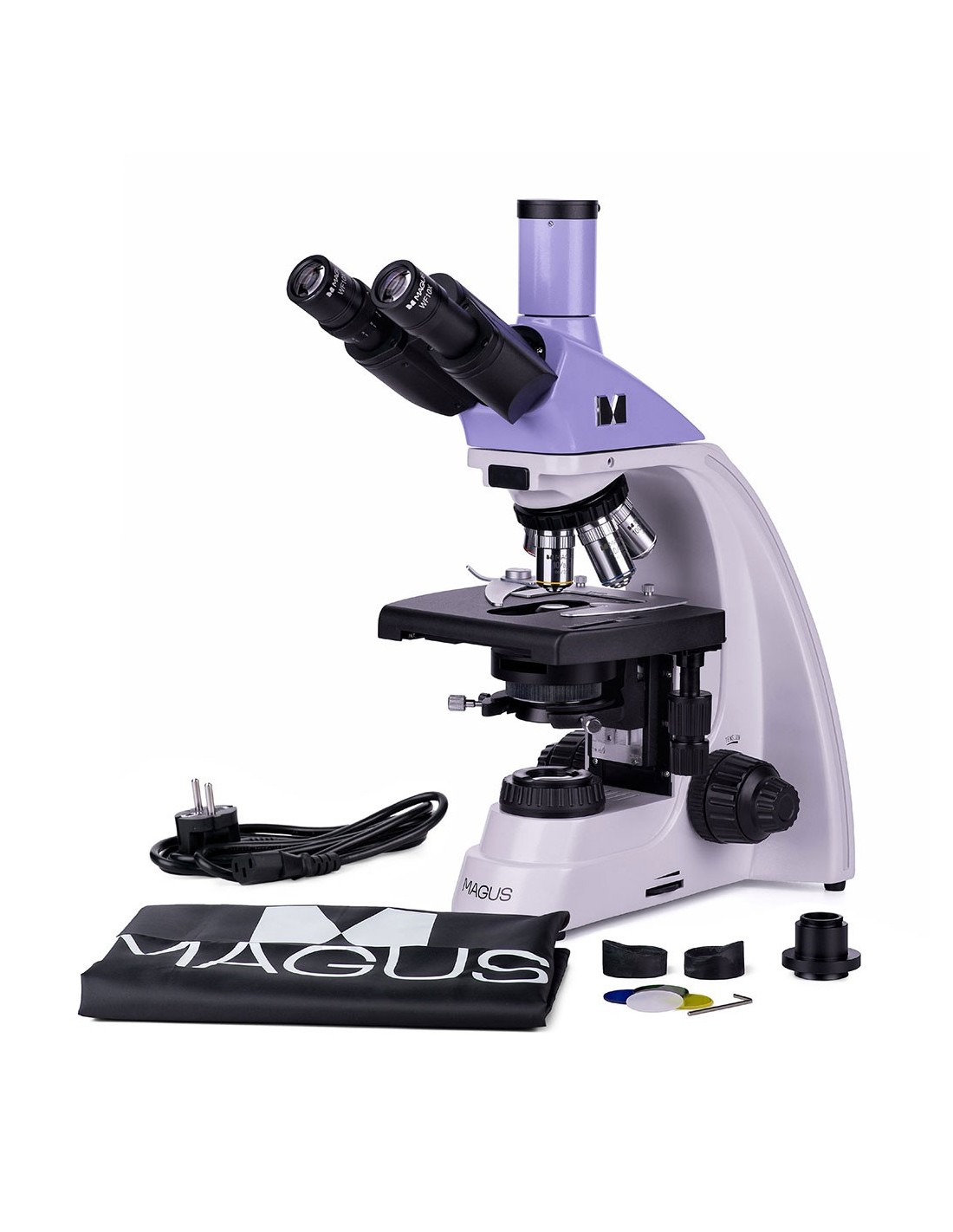
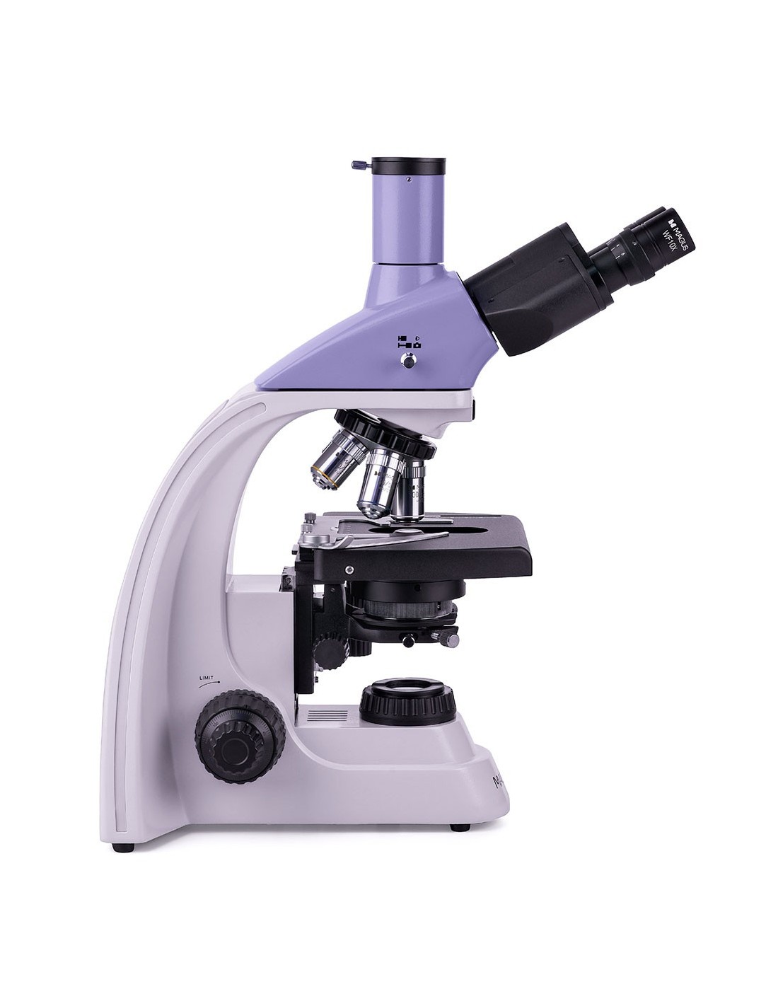
Magus Bio 230T is a trinocular biological microscope for professional observations in laboratories and research centers in the medical, pharmaceutical, forensic, biotechnological fields, etc. It is suitable for the observation of semi-transparent biological preparations and naturally transparent samples: thin sections and smears. With the use of achromatic objectives and a halogen lamp, the analysis of the samples is carried out using a transmitted light (bright field) microscopy technique. The design of the microscope offers the possibility of installing additional accessories that allow you to use other microscopy techniques: dark field, phase contrast and polarized light.
Digital camera
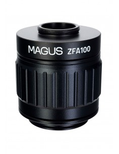
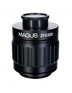
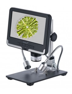
MAGUS CHD10 is the camera with HDMI interface. The camera is equipped with a 2 MP sensor and produces realistic Full HD images at a resolution of 1920x1080 pixels.
It is a standalone camera that does not require a connection to the computer or the installation of additional software. You can connect the camera directly to a TV, monitor or projector to display an image.
The HDMI interface allows for fast and stable speed transfer from the camera to the external screen. Video is recorded at 60 fps.
The camera combines high FPS rate and high HDMI bandwidth. The videos therefore appear brilliant without freezing or skipping between frames. At maximum resolution, the image is very detailed, moving objects are visible without errors, and the moving object is displayed without delays.
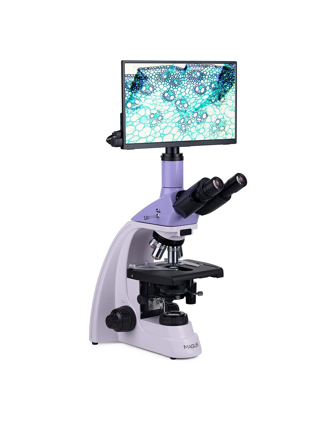
Monitor
MAGUS MCD monitors work in combination with MAGUS CHD cameras to form a complete imaging system for MAGUS microscopes.
The connection to the camera is via its HDMI interface. The monitor displays images in real time. Its IPS LCD display has a wide viewing angle, high brightness and good contrast. When looking at the screen even from a wide angle, the image is not distorted. The screen size is 13.3''.
The monitor can be installed on a horizontal surface using a foldable stand or mounted directly on a camera or tripod.
Optics
Optics
strong>
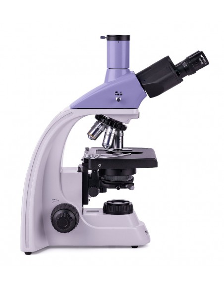
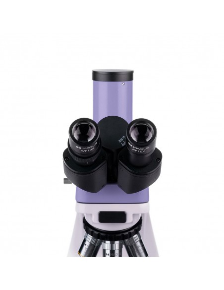
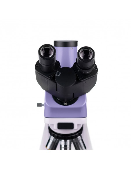
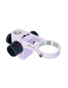
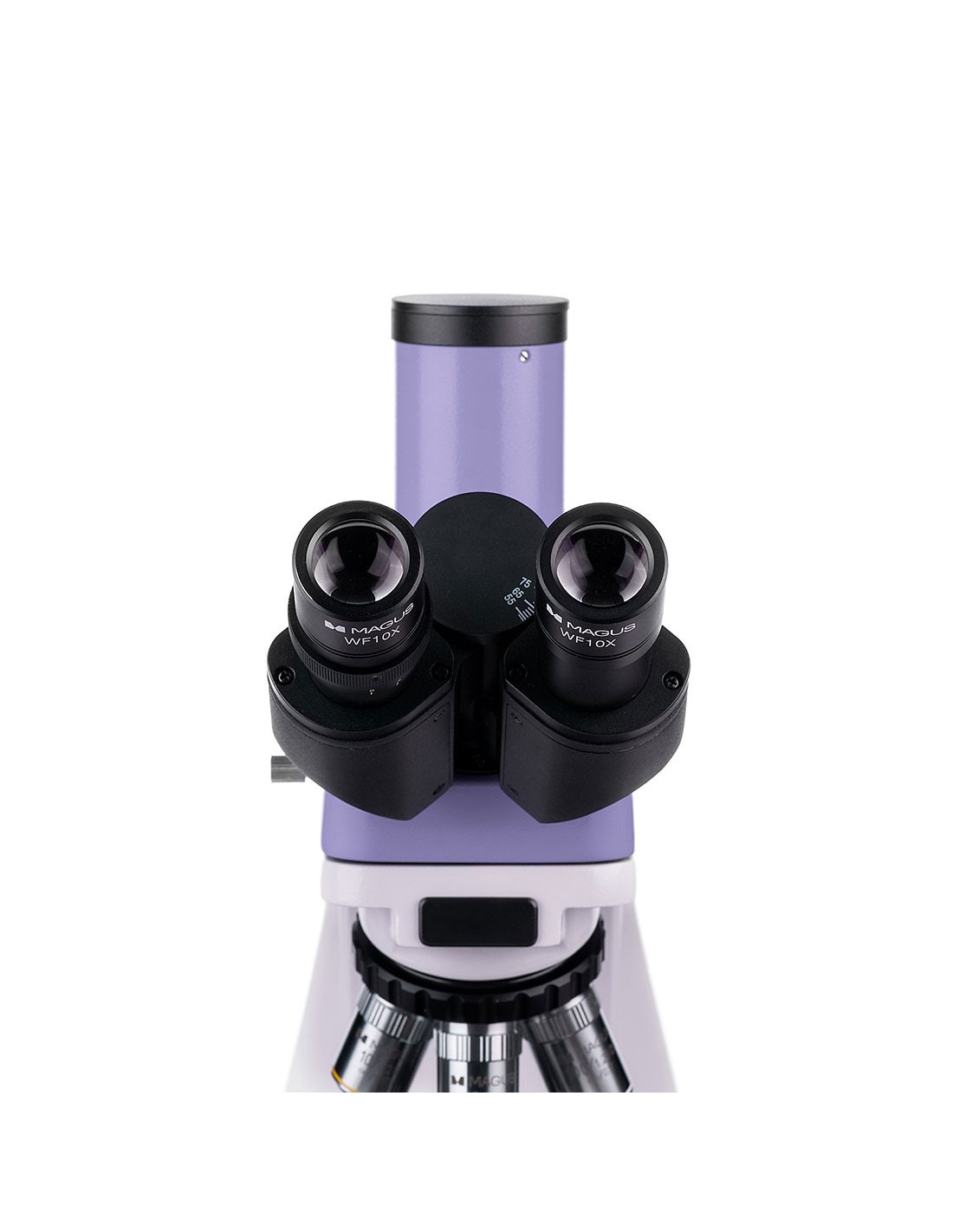
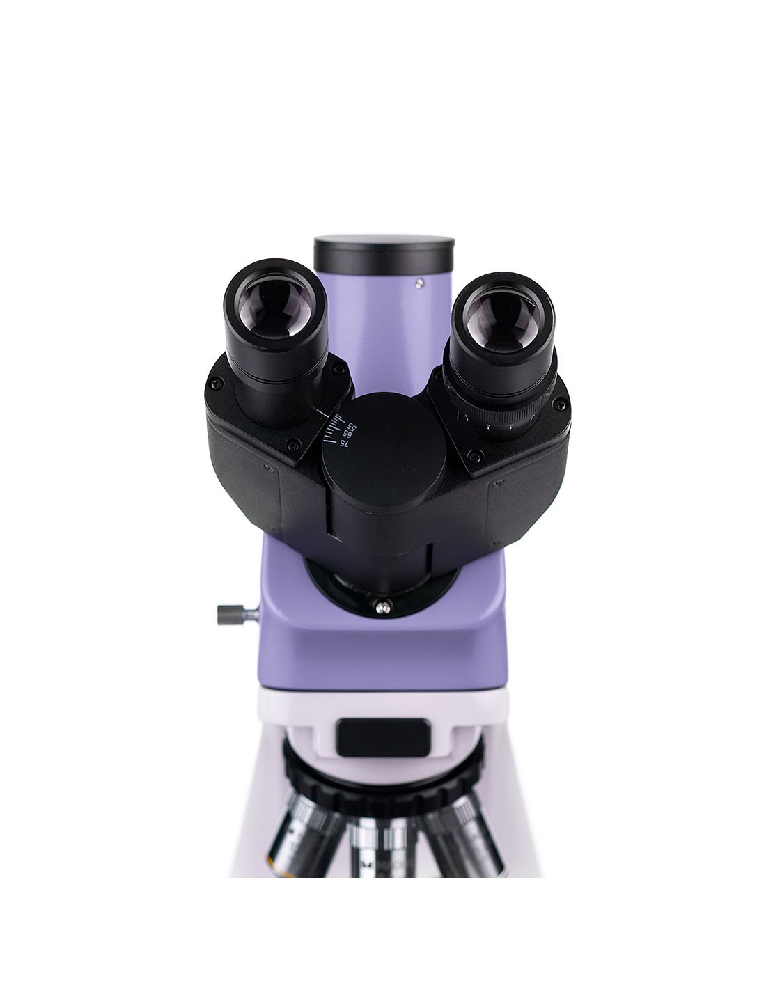
The trinocular head is equipped with a vertical tube for housing a digital camera. The camera (not included) is an excellent solution for creating a digital archive of your observations and can be used to transmit images to an external monitor. To adapt the optical tubes to the observer's height, they can be rotated 360°. The left tube is equipped with a diopter adjustment ring.
The nosepiece is oriented towards the inside of the instrument, therefore, the space above the stage remains free. Five lenses can be mounted on the nosepiece at the same time (4 are included in the package and a fifth slot is left empty for an additional lens). In their basic configuration, infinity-corrected objectives provide magnification from 40x to 1000x. With the use of additional eyepieces, the magnification can be increased up to 1500x, 1600x or 2000x.
Illumination
The halogen lamp produces a warm colored lighting, which has less impact on eye strain and is ideal for those who work long hours at the microscope. The lamp is 30 W, therefore the brightness and contrast of the transmitted image are high with any type of objective or observation technique.
The Abbe condenser is adjustable in height and can be centered. Illumination is controlled by adjusting the field diaphragm and iris diaphragm; Furthermore, Köhler illumination can be set up for improved image clarity. The condenser features a slot to accommodate a dark field or phase contrast slider, making switching between various microscopy techniques quicker and easier.
Stage and mechanism focusing
There is no positioning rack for the stage. This is an advantage in the design of the mechanical stage shifter, as it makes it easier to operate the microscope. The sample can be moved smoothly across the stage. The mechanical insert is removable (it can be removed to perform a manual scan).
It is possible to adjust the coarse and fine focus (coarse and fine screw); The movement of the focus knob is smooth, simple, and requires no effort. The coarse focus mechanism is equipped with a fixing knob and the tension of the coarse screw can be adjusted. The focus knobs are coaxial and positioned to ensure maximum comfort during long hours of observation: they are, in fact, located on the base of the microscope, so that the observer can comfortably rest his hands on the table.
Accessories
To maximize the capabilities of the Magus Bio 230T microscope, it is possible to enhance it with some accessories. These may include eyepieces and objectives, digital cameras and calibration slides, dark field condensers, polarizers, and instruments for phase contrast observations.
Key Microscope Features:
Key Features of the Microscope:
strong>
- Trinocular head with vertical tube for housing a digital camera
- The 360° rotation allows the instrument to be adapted to the height of the observer
- li>
- Infinity corrected achromatic objectives, nosepiece nosepiece for 5 objectives
- Diopter adjustment ring on the left optical tube
- 30 W halogen lamp powered by AC mains, Abbe condenser with slot for dark field slider or for phase contrast, field diaphragm
- Coaxial knobs for coarse and fine focusing (macro- and micrometric screw), fixing knob and adjustment knob tension adjustment for the coarse screw
- Optional accessories to improve the performance of the microscope
Key camera features:
- Standalone camera operation without PC connection or software installation
- HDMI interface for stable and fast data transfer
- 1920x1080 pixel resolution, the ideal choice for viewing of images on a Full HD monitor
- 60 fps for observing moving samples, video recording and sample movement without delays or stutters
- The color back-illuminated SONY Starvis CMOS sensor It offers images with low noise and high light sensitivity even in low light conditions. You can get sharper, brighter and more saturated color images
- Software for photo and video recording and editing, external display, linear and angular measurements
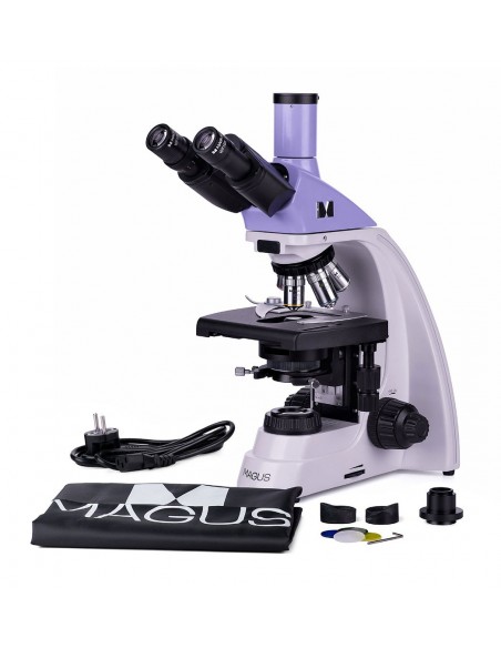
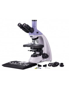
The package contains:
- MAGUS CHD10 digital camera (digital camera, HDMI cable (1.5 m), mouse with USB, 32 GB SD memory card, power adapter AC 12 V/1 A (Euro), user manual and warranty certificate)
- MAGUS MCD20 LCD monitor
- Base with power input, light source for transmitted light observations, mechanism focusing mechanism, stage, condenser attachment and nosepiece revolver
- Abbe condenser
- Trinocular head
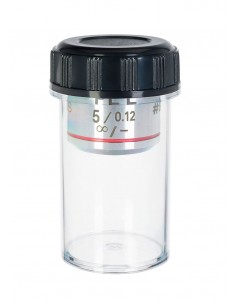
- Achromatic infinity objective: 4x/0.10
- Achromatic objective at infinity: 10x/0.25
- Achromatic objective at infinity: 40x/0.65 (with spring mechanism)
- Achromatic objective to infinity: 100x/1.25 oil (with spring mechanism)
- 10x/18 mm eyepiece with long eye relief (2 pcs.)
- Eyecup (2 pcs.) )
- Filter (4 pcs.)
- C-mount adapter
- Vial of immersion oil
- AC power cord
- Dust cover
- User manual and warranty card
Available upon request:
- 10x/20 mm eyepiece with graduated scale
- 15x/11 mm eyepiece (2 pcs.)
- 16x/11 mm eyepiece (2 pcs.)
- Eyepiece 20x/11 mm (2 pcs.)
- Achromatic objective at infinity: 20x/0.40
- Planachromatic objective at infinity: 20х/0.40
- Infinity plan achromatic objective: 60x/0.80 (spring loaded)
- Phase contrast device
- Phase contrast slider
- Dark field condenser with NA 0.9
- Oil dark field condenser with NA 1.36–1.25
- Dark field slider
- Polarization instruments
- Calibration glass
| Brand | MAGUS |
| Warranty | 5 лет |
| EAN | 5905555017952 |
| Shipping weight | 12.65 kg |
| Delimiter | MAGUS Bio 230T biological microscope |
| === Delimiter === | Specifications of the microscope |
| Type | biological, light/optical, digital |
| Tested | trinocular |
| Vertical tube | Gemel head (Siedentopf type, with 360° rotation) |
| Tilting angle of the head | 30° |
| Magnification, x | 40–1000 in the basic configuration (*optional : 40–1500/1600/2000) |
| Magnifying range | 800x to 1280x |
| Eyepiece tube diameter, mm | 23.2 |
| Eyepieces | 10х/18 mm, eye relief: 10 mm (*optional: 10x/20 mm with scale, 15x/11 mm, 16x/11 mm; 20x/11 mm) |
| Infinity corrected achromatic objectives: 4x/0.10; 10x/0.25; 40xs/0.65; 100xs/1.25 (oil); parfocal distance 45 mm (*optional: 20x/0.40; 60xs/0.80) |
| Nosepiece nosepiece | for 5 objectives |
| Working distance, mm | 18.89 (4x); 5.95 (10x); 0.775 (40xs); 0.36 (100xs); 2.61/8.80 (20x); 0.46 (60хs) |
| Interpupillary distance, mm | 48 — 75 |
| Stage, mm | 180x150 |
| Range of movement of the stage, mm | 75/50 |
| Stage features | two-axis mechanical translator, without positioning rack |
| Eyepiece diopter adjustment, diopters | ±5 (on left barrel) |
| Condenser | Abbe condenser, NA 1.25, adjustable centering, adjustable height, adjustable aperture diaphragm, with slot for dark field slider and phase contrast slider, dovetail mount |
| Diaphragm | adjustable aperture diaphragm, field diaphragm with adjustable iris |
| Focus | coaxial, coarse focus (21 mm, 39.8 mm/turn, with fixing knob and focus adjustment knob tension of the micrometer screw) and fine focus (0.002 mm micrometer screw) |
| Halogen |
| Brightness adjustment | yes |
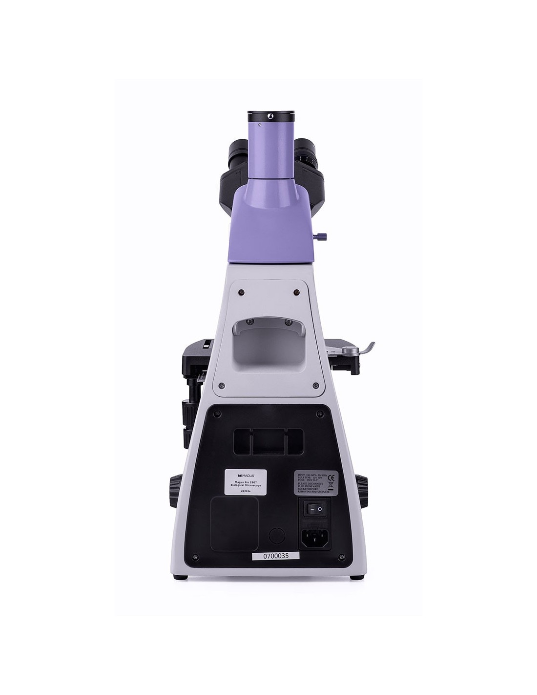
| Power supply | 220±22 V, 50 Hz, AC mains |
| Type of light source | 12 V/30 W halogen lamp, G4 socket |
| Optical filters |
| Optical filters | yes |
| Operating temperature range, °C | 5 — 35 |
| Possibility to connect additional equipment | phase contrast devices (condenser and objectives), dark field condenser (dry or oil immersion), polarization instruments (polarizer and analyzer) |
| User level | expert users, professionals |
| Assembly and installation level of difficulty | complicated |
| Application | laboratory/medical |
| Lighting position | lower |
| Search method | bright field |
| Set with pencil case/case/bag | dust cover |
| Weight, kg | 8 |
| Dimensions, mm< /td> | 200x436x400 |
| === Delimiter === | Digital camera |
| Sensor | SONY Starvis CMOS |
| Color/monochrome | color |
| Megapixel | 2 |
| Maximum resolution, px | 1920x1080 |
| 1/2.8" (5.57x3.13mm) |
| Pixel size, µm | 2.9 x2.9 |
| Interface connectors | HDMI 1.4 |
| Memory card |
| Memory card |
| td> | SD up to 32 GB |
| Possibility to connect additional equipment | mouse with USB |
| Light sensitivity | 1300 mV at 1/30 s |
| Exposure time | 0.04 ms – 1000 ms |
| Video recording | yes |
| Installation location | pipe trinocular, optical tube instead of eyepiece |
| Image format | *.jpg |
| Video format |
| Video format |
| td> | *.h264, *.mp4 |
| Spectral range, nm | 380-650 (integrated IR filter) |
| Shutter type | ERS (electronic curtain shutter) |
| Software | HDMI: integrated |
| System requirements | does not require a computer connection |
| Mount type< /td> | C-mount |
| Body | robust aluminium |
| Camera power supply | AC adapter 12 V, 1 A |
| AC power adapter specifications | input: AC voltage 100–240 V, 50 /60 Hz, output: DC voltage 12 V/1 A |
| Camera operating temperature range, °C | -10 — 50 |
| Humidity operating range, % | 30 — 80 |
| === Delimiter === | Monitor |
| Matrix type | IPS |
| Display diagonal, inches | 13.3 |
| Display resolution, px | 1920x1080 (Full HD) |
| Format | 16:9 |
| Brightness, cd/m² | 400 |
| 16.7 m |
| Contrast | 1000:1 |
| Horizontal/vertical viewing angle, ° | 178/178 |
| Dimensions of the visible area of the screen (WxH), mm | 295x165 |
| Pixel pitch (WxH), mm | 0.154x0.154 |
| Matrix type backlight | LED |
| LED backlight life, h | 50000 |
| Interface | HDMI |
| Operating temperature range, °C | -15 — 55 |
| Humidity operating range, % | 10 — 90 |
| Power Supply | AC 110–220 V, DC 5–12 V/1 A (Type C) |
| Energy consumption, W | 12 (max.) |


