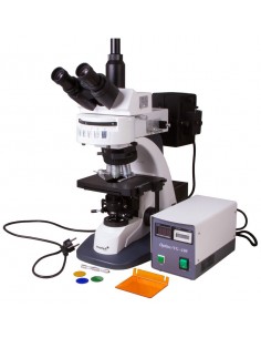
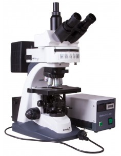
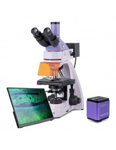
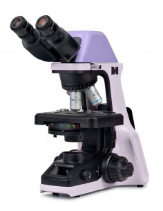
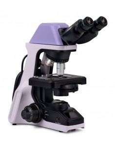
The Levenhuk MED D45T trinocular microscope is equipped with a 16MP digital camera that allows you to take photos and record videos in high resolution. The microscope is suitable for phase contrast observations as well as bright and dark field observations. It is the perfect resource for a clinical diagnostic center, biochemical laboratory or microbiological laboratory. The microscope has a Köhler illumination adjustment; all observations are carried out with a magnification of 40x to 1000x.
The advantages of the phase contrast method
It is worth underlining that this optical instrument It also allows phase contrast microscopy. This method improves the contrast and sharpness of images of transparent and semi-transparent samples to a level achievable by the standard method only by treating the samples with dye. This is why phase contrast microscopy is essential for studying samples in vivo in their natural conditions, as the staining process would, in fact, cause cell death. This method can be applied to the analysis of drinking water and parasites, to cytological, hematological analyzes and other research fields.
Planachromatic optics with infinity correction p>
The Levenhuk MED 45 series microscopes are equipped with an infinity corrected optical system, used in professional and high-end microscopes. This system includes planachromatic objectives and allows the transmission of clear, high-contrast images with an excellent level of planarity.
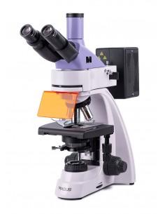
One of the most important characteristics of the infinity corrected optical system is that it allows the installation of further parts in the optical path between the objective lens and the eyepiece. Additional parts include epi-fluorescence polarizer and devices. Overall, the modular design and ease of use make Levenhuk MED 45 microscopes optimal tools to use in various fields of microscopy, for work in hematology, histology, microbiology laboratories and much more.
16M digital camera for capturing photos and videos
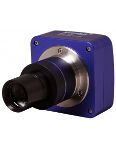
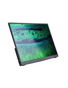
The trinocular head includes the 30-degree inclined binocular viewing part and the vertical eyepiece tube in which the digital camera is placed. The kit includes a camera with a 16 MP sensor. It can be used to transmit images from the lens to an external screen as well as take photos of specimens and record videos of studies. A camera transmits the image to the screen of a connected computer. This makes laboratory work simpler and more comfortable, as well as reducing strain on the eyes, as there is no need to observe through eyepieces. The camera is connected to the PC via a USB cable included in the kit. Additionally, image editing software can be installed from the disk. The software allows you to change image size, brightness and contrast, exposure time and white balance, calibrate the camera and objective lenses, as well as measure specimens and structures (various units of measurement available).
Optical Capabilities
Optical accessories include planachromatic phase contrast objective lenses of various magnifications and 10x wide-field eyepieces with diopter adjustment. The 40x and 100x objective lenses feature spring-loaded mounts that protect the optics from accidental damage. The optical system produces clear, high-contrast images without chromatic aberration. A 100x objective is for oil immersion. All accessories are made of optical glass with antifungal coating.
Work comfortably with slides
The stage with double coordinates has a mechanical translator that facilitates positioning timely and accurate sampling of samples under the objective. Coarse and fine focus are available. Köhler lighting control is available. An LED light sits at the bottom and has a brightness adjustment. The phase contrast condenser can be used for dark field studies with the “dry” method.
The light is powered by an AC power supply.
Features:
- Magnification range of the trinocular head from 40x to 1000x
- Planachromatic optical system for infinity corrected phase contrast
- Wide field eyepieces with diopter adjustment
- Dark field phase contrast condenser
- Bottom LED light with brightness adjustment
- Köhler illumination available
- 16MP digital camera is included
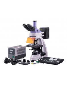
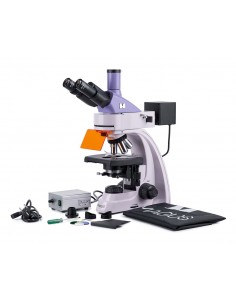
Kit includes:
- Microscope base with stand
- 360° rotatable trinocular head
- Planachromatic objective lenses for phase contrast, infinity corrected: 4X, 10x, 40xs, 100xs (oil) with antifungal coating
- Wide field eyepieces: WF10x/22 mm with antifungal coating (2 pcs.)
- Phase contrast device (dark field)
- Filters: blue, green, yellow
- Bottle of immersion oil
- Fuses (2 pcs.)
- Microscope power cord
- Dust cover
- 16MP digital camera
- Camera adapter
- Camera mount
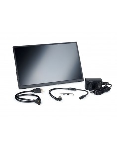
- USB cable to connect and charge the camera
- CD with software and drivers
- User manual and lifetime warranty
Attention:
for information regarding the correct mains voltage, consult the specifications table. Never attempt to connect a 110V device to a 220V power outlet and vice versa without using a voltage converter. Please note that mains voltage is 220–240 V in most European countries and 110 V in the United States and Canada.
Some things you can observe under a microscope:
The Levenhuk MED D45T Digital Trinocular Microscope is compatible with Levenhuk digital cameras (sold separately). Levenhuk cameras are installed on the optical tube, in place of an eyepiece.
| Brand | Levenhuk Inc., USA |
| Lifetime guarantee | |
| EAN | 5905555005089 |
| Package size (LxWxH) | 50x43x28 cm |
| Shipping weight | 10.5 kg |
| Type | digital, light/optical, biological |
| Tested | trinocular |
| Optics material | optical glass with anti-mold coating |
| Vertical tube | rotatable 360° |
| Head tilt angle | 30° |
| Magnification, x | 40 — 1000 |
| Magnifying range | 800x to 1280x |
| Diameter eyepiece tube, mm | 30 mm (binocular head), 23.2 mm (third vertical tube) |
| Eyepieces | WF10x /22 mm, wide field with diopter adjustment (2 pcs.) |
| Infinity-corrected planachromatic phase contrast objectives: 4x, 10x, 40xs, 100xs (oil immersion) |
| Nosepiece nosepiece | for 5 objectives |
| Interpupillary distance , mm | 48 — 75 |
| Coffee table, mm | 180x150 |
| Range of movement of the stage, mm | 75/50 |
| Stage characteristics | double mechanical layer |
| Diopter adjustment of the eyepiece, diopters | ±5 |
| Condenser | phase contrast ( with dark field) |
| Focus | coaxial, coarse (0.5 mm) and fine (0.002 mm), with rack and pinion |
| Body | metal |
| Lighting | LED |
| Brightness adjustment | yes |
| Power supply | 100–240V |
| Type of light source | 5 W, 85–230 V AC |
| Optical filters | blue, green, yellow |
| Extra elements | collector, Köhler lighting |
| User level | expert users, professionals |
| Assembly and installation level of difficulty | complicated |
| Application | laboratory/medical |
| Lower lighting position | |
| Method of research | bright field, dark field, phase contrast microscope |
| Digital camera included | yes |
| Set with case/case/bag | dust cover |
| Maximum resolution | 4632x3488< /td> |
| Megapixel | 16 |
| Sensor element | 1/2,33 " |
| Pixel size, µm | 1.335x1.335 |
| Video recording | yes |
| Image format | *.jpg, *.bmp, *.png, *.tif, etc. |
| Video format | recording: *.wmv, *.avi, *.h264 (Windows 8 and later), *h265 (Windows 10 and later) |
| Spectral range, nm | 380-650 |
| White balance | manual, automatic |
| Exposure control | 0.2–2000 μs |
| Frame rate | 2@4632x3488
8@2320x1740
11@1536x1160 |
| Position of use | the third eyepiece tube of the 23.2 mm microscope mm |
| Software, drivers | LevenhukLite |
| Programmable options | size image, brightness, shutter speed |
| Output | USB 2.0 |
| System requirements |
| System requirements |
| td> | Windows 8/10/11 (32-bit and 64-bit), Mac OS |
| Camera power supply | via USB cable |


